Lifeasible's experimental team has extensive experience in exosome identification and can analyze exosome properties comprehensively. Exosome identification is designed to assess the effectiveness of an investigator's exosome isolation and purification by determining the presence, number, and integrity of exosomes in enriched exosome samples through different experiments. However, a single identification does not accurately identify the presence, quantity, and integrity of exosomes and needs to be illustrated by the following three experimental data together.
 Figure 1. Exosome identification. (X. Wu et al., 2019)
With a resolution of 0.1 - 0.2 nm, transmission electron microscopy can magnify the observed object millions of times, which is ideal for the ultrastructural observation of exosomal bilayer vesicles. Lifeasible's team is engaged in exosome research and has accumulated rich experience in processing and identifying exosome electron microscopy samples, providing exosome researchers with high-quality exosome TEM detection services.
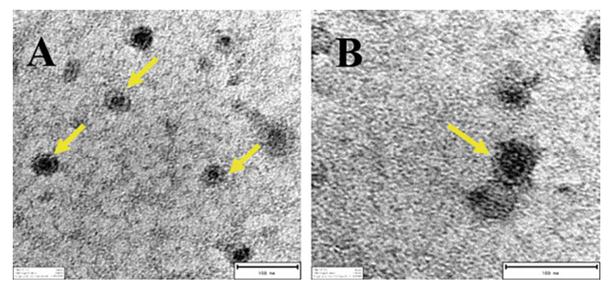 Figure 2. Transmission electron microscopy results. Yellow arrows: exosomes. (X. Wu et al., 2019)
Nanoparticle Tracking Analysis (NTA) technique is a novel nanoparticle characterization method which obtains the scattered light pattern of each nanoparticle by optical microscopy and tracks and analyzes the Brownian motion of each particle to calculate the hydrodynamic diameter and concentration of nanoparticles. The NTA technique allows for rapid and accurate analysis of exosome particle size distribution concentrations, providing strong evidence for the presence of exosomes. Compared to other characterization methods, the NTA technique is simpler to handle, better able to guarantee the original state of exosomes, and faster to detect.
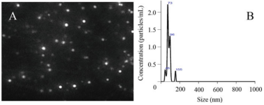 Figure 3. Exosome size distribution. (Wu et al., 2019)
Western Blot is a commonly used assay in molecular biology, biochemistry, and immunogenetics. The basic principle is to separate the proteins in the cell, or tissue samples by polyacrylamide gel electrophoresis, the target proteins are bound to the specific primary antibody after membrane transfer and closure then react with the horseradish peroxidase (HRP)-labeled secondary antibody. Analyze the position of the exposed bands and grayscale analysis to know the expression of the target proteins in the samples. This method combines the high specificity and sensitivity of gel electrophoresis and solid-phase immunoassay techniques, allowing both definitive and semi-quantitative analysis of the target protein.
Lifeasible can provide a wide range of exosome identification services, offering exosome transmission electron microscopy identification results, accurate exosome particle size analysis, and exosome surface marker expression identification. If your product needs to be identified, contact us, and we can provide solutions for the above identification methods to help you complete your project as soon as possible.
Lifeasible will provide you with a complete set of services from sample preparation, sample purification, and sample identification to data analysis.
Experienced team with expertise in cutting-edge fields to provide our customers' solutions.
A strong bioinformatics team is available to meet the needs of our clients for in-depth data analysis.
Reference

Why Do Plants Blush When They Are Hungry?
April 26, 2024

STU-CRISPR System Improves Plant Genome Editing Efficiency
April 19, 2024