Fluorescence resonance energy transfer (FRET) is a physical phenomenon which relies on the distance-dependent energy transfer. The occurrence of FRET requires a donor molecule and an acceptor molecule. When the two molecules are in close proximity (less than 10 nm), the transfer of energy lead to the reduction of the donor’s fluorescence intensity and the increase of the acceptor’s emission intensity, and the donor and acceptor molecules are called a FRET pair.
Fluorescent labels, such as YFP, CFP, GFP and RFP, can form FRET pairs with each other, as long as the emission spectrum of the donor overlaps with the excitation spectrum of the acceptor. Based on this principle, FRET can be combined with high-resolution microscopy imaging to detect protein-protein interactions in living cells. Specifically, two proteins of interest can be tagged with two fluorophores that can form a FRET pair (eg. CFP and YFP), respectively. If there is no interaction between these two proteins and they are distributed beyond certain distance, the two fluorophores will show normal fluorescence behaviors. While if the two proteins interact with each other, the excitation energy of CFP will be transferred to YFP in a non-radiative fashion, which will lead to an emission of yellow fluorescence only (Figure 1).
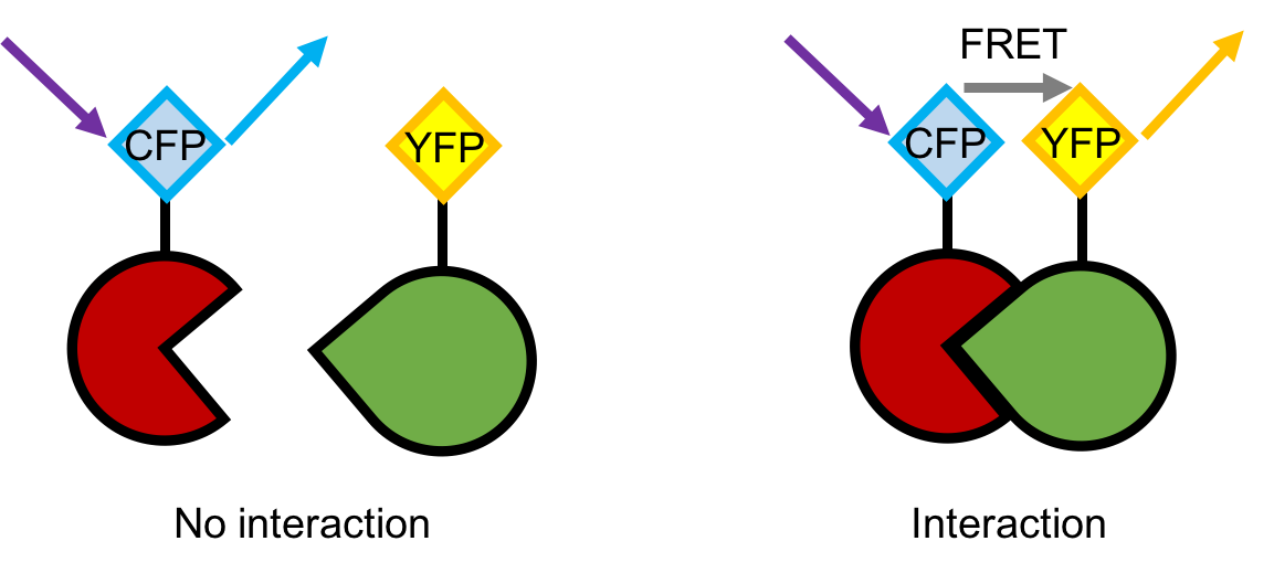 Figure 1. A schematic description of the principle of FRET microscopy imaging.
As an advanced technology for the detection of protein-protein interactions, FRET microscopy imaging has the following advantages
Lifeasible is a leading plant biotechnology company. With years of experience in plant molecular biology, plant biochemistry and plant cell biology, we provide high-quality FRET microscopy imaging services in the plant system, with the most advanced microscopy techniques, flexible choices of fluorophore pairs and professional image analysis platforms. Moreover, our featured one-stop service covers every single step of your project, including:
Welcome to contact us for questions, inquires or collaborations.
Get Latest Lifeasible News and Updates Directly to Your Inbox