Fluorescent microscopy is a cornerstone of modern cell biology. However, fluorescent labeling can result in the loss of the surrounding cellular environment, and certain fluorescent stains may affect the natural structure of cells or organelles. Therefore, when you want to understand the ultrastructure inside tissues and cells, cryo-electron microscopic (Cryo-EM) imaging is the way to go. Cryo-EM is an advanced imaging technique widely used to explore the structure of subcellular organelles.
As a professional bioimaging company, Lifeasible has built up a cutting-edge cryo-electron microscopy imaging technology platform. We provide one-stop cryo-electron microscopic imaging services for botanists worldwide to help them elucidate the 3D structure of subcellular organelles and protein complexes, as well as organelle interactions.
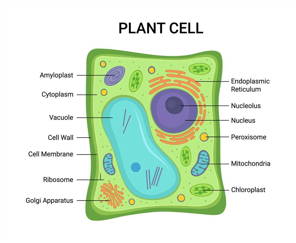
Cryo-EM can help us to observe the three-dimensional structure of plant subcellular organelles in different states, as well as all complex conformations, structures, and modified forms of proteins in subcellular organelles. At the same time, Cryo-EM only requires a small sample and does not require crystallization of biological samples, avoiding the impact of purity and heterogeneity issues on research.
Depending on your needs, our Cryo-EM technology platform can provide you with several imaging services, such as cryo-transmission electron microscopy, cryo-scanning electron microscopy, cryo-electron tomography, and cryo-etching electron microscopy.
Cryo-TEM usually adds a sample freezing device to an ordinary transmission electron microscope to observe temperature-sensitive samples such as proteins and biological sections. By freezing the sample, the damage to the sample by the electron beam can be reduced, and the deformation of the sample can be reduced, thereby obtaining a more realistic sample morphology.
Cryo-SEM technique is a fast, reliable, and effective method to overcome the problem of sample water content. Cryo-SEM is the most effective way to prevent moisture loss from the sample, and it can be used in any vacuum.
Cryo-ET images at nanoscale resolution, producing three-dimensional (3D) images from a series of tilted Cryo-EM images. It is commonly used for many structures inside cells, such as subcellular organelles.
Freeze-etching technology is used for microstructural studies in fields such as cell biology. The sample is frozen so that the microstructure is close to that of a living organism; after freeze-etching, the microstructure of the different cleavage surfaces can be observed, allowing the study of the membrane structure and inclusions within the cell.
With the rapid development of science and technology, fluorescence microscopy imaging is becoming more and more widely used in the biomedical field. Our imaging technology platform offers the following types of fluorescence microscopes.
Lifeasible is committed to providing robust technical support for observing plant subcellular organelles. If you are interested in our plant subcellular organelle cryo-electron microscopy imaging services, please feel free to contact us. We look forward to working with you.
Get Latest Lifeasible News and Updates Directly to Your Inbox