Plant subcellular organelles are essential units of the cell; their morphological structure and dynamics directly reflect the cell's physiological state, and each subcellular organelle has a unique structure. The depth of structural analysis of subcellular organelles often depends on the development of imaging techniques. In recent years, the field of subcellular imaging has made great progress with developing new techniques, and the structural analysis of subcellular organelles has entered a new phase.
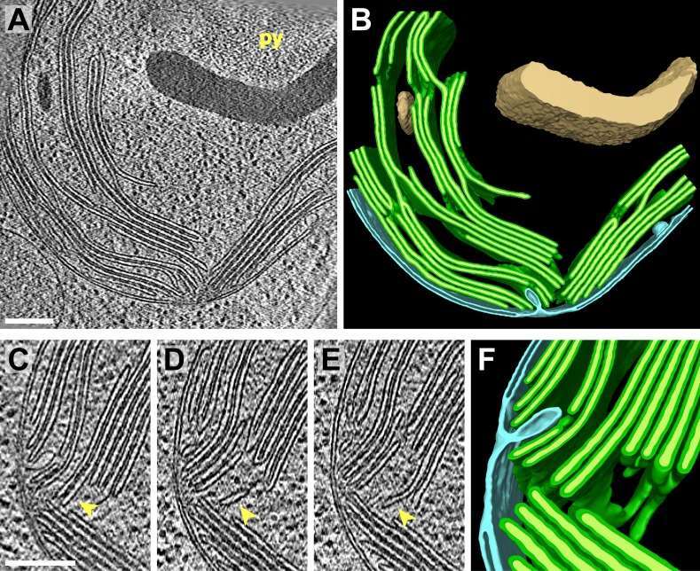 Figure 1. Structure of chloroplasts of Chlamydomonas reinhardtii as revealed by in situ cryo-electron tomography. (Engel, B. D, et al. 2014)
Lifeasible has been working in bioimaging for many years and has extensive experience in plant subcellular structure analysis. We have a wide range of advanced imaging machines and a professional subcellular structure analysis team to provide quality plant subcellular organelles structure analysis services for clients engaged in cell biology research.
For different needs and research purposes, we will provide the most suitable technical support solutions. We can offer our customers three imaging techniques, including cryo-electron microscopy, fluorescence imaging, and super-resolution microscopy; the main ones are as follows.
The advantage of Cryo-TEM is mainly in the high accelerating voltage, which allows electrons to penetrate thick samples. The technique is suitable for structural biology and structure determination, including the study of subcellular structures.
Cryo-SEM allows direct observation of liquid and semi-liquid samples; it avoids cell distortion and has the advantage of being easy and fast to prepare. It is very suitable for imaging and analysis of subcellular organelles structure.
Cryo-ET is a label-free cryo-imaging technique that provides 3D datasets of organelles and protein complexes at nanometer resolution and is suitable for the structural analysis of cellular organelles.
The use of specific fluorescent dye markers to target different organelles, followed by structural analysis of the target organelle by fluorescence microscopy. This method is suitable for targeted analysis of a particular organelle.
SIM technique does not require special fluorescent markers and is suitable for live cell imaging, especially for long-duration imaging of live cells.
STED microscopy takes advantage of the stimulated emission effect and the non-linear effect to reduce the fluorescence area and thus increase the resolution. Typical STED microscopy systems have a resolution of approximately 50 nm.
The SMLM technique does not require much in the way of experimental equipment but can achieve high levels of resolution (around 20 nm or even less than 10 nm). Still, it requires special fluorescent molecules for labeling and has a low temporal resolution.

A full understanding of the complex structure of subcellular organelles requires nanoscale 3D reconstruction, and Lifeasible provides customers with solutions to reveal the 3D structure of organelles. We can help our clients understand the structure of organelles and the dynamics of organelle structure in specific situations. If you are interested, please feel free to contact us.
Reference
Get Latest Lifeasible News and Updates Directly to Your Inbox

