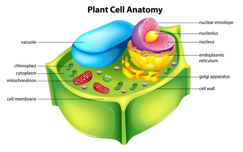
Plant subcellular organelles include the nucleus, endoplasmic reticulum, Golgi apparatus, chloroplasts, mitochondria, vesicles, centrosomes, and peroxisomes, which are the basic structures that sustain the normal life activities of plants. The plastids and vesicles can be distinguished under the light microscope, while the other organelles are usually observed with an electron microscope. With advances in biotechnology and the demand for higher-quality imaging of plant subcellular organelles, fluorescence imaging techniques have been widely used to image organelles. Changes in organelle structure, function, and interactions can be visualized by fluorescence microscopy and fluorescent dyes.
Lifeasible, as a globally renowned biotechnology company, has always been committed to a human-centered service philosophy. Based on our advanced imaging technology platform, we can provide our customers with high-quality fluorescence imaging services of plant subcellular organelles, which can help them understand organelle positioning, organelle structure, function and interactions in detail, etc.
Organelles play a key role in cellular function and the use of appropriate organelle dyes, probes, or specific antibodies for the detection of specific organelles is critical in cell and tissue fluorescence imaging. Our one-stop shop for plant subcellular organelle fluorescence imaging services will try to meet your needs from designing and synthesizing specific fluorescent probes to analyzing imaging results. Our services include but are not limited to, the following items.
With the rapid development of science and technology, fluorescence microscopy imaging is becoming more and more widely used in the biomedical field. Our imaging technology platform offers the following types of fluorescence microscopes.
The most common fluorescent materials used for fluorescence imaging include organic fluorescent dye molecules, fluorescent proteins, and fluorescent nanomaterials. These probes are essential for long-term tracking of biological processes, low autofluorescence tissue imaging, and time-resolved fluorescence imaging. We offer optimized fluorescent dyes with low cytotoxicity, high photostability, near-infrared (NIR) emission, two-photon excitation, and long fluorescence lifetimes.

Lifeasible's fluorescent imaging services label and identify organelles of interest to you in fixed and live cells using specific fluorescent dyes. If you are interested, have questions, or would like to collaborate, please contact us directly.
Get Latest Lifeasible News and Updates Directly to Your Inbox

