Detection of Programmed Cell Death in Plant Cells
Programmed cell death (PCD), known as apoptosis, is a genetically regulated biological phenomenon that occurs in cells under specific physiological or pathological conditions. PCD is an essential component of plant growth and development, and it occurs throughout embryonic development, cell differentiation, and organ formation. At the same time, PCD is also a basic mechanism for plants to resist biotic or abiotic stresses. It plays a vital role in the process of plant disease resistance or stress tolerance. The detection and study of programmed cell death in plants can not only reveal the internal law of aging and death but also provide the basis and technology for regulating growth and development.
As an expert in plant-programmed cell death detection, Lifeasible provides a variety of effective techniques for the detection of programmed cell death. In addition to morphological observation by optical microscope and electron microscope, we also utilize molecular biology, cell biology, and other related techniques in the detection of plant PCD, which greatly improves the accuracy and efficiency of the detection.
Methods for the detection of PCD in plant cells
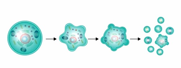
When plant cells develop PCD, typical morphological changes include cytoplasmic and nuclear crumpling, highly condensed and marginalized chromatin, endoplasmic reticulum expansion, mitochondrial swelling, and vesicle rupture. We can observe plant cells undergoing PCD using light, fluorescence, laser confocal, and electron microscopy. The following are some of the morphological detection methods.
- Light microscopy detection. After plant tissue samples are stained and treated with specific dyes, morphological changes such as cell volume reduction, coalescence, margination, and apoptotic vesicle formation can be observed by light microscopy to quickly determine the production of PCD. At present, the optical microscopy method can only be used for the preliminary discrimination of PCD in plant cells and should be combined with other means of detecting PCD.
- Fluorescence microscopy detection. Unlike necrotic, PCD and normal cells have intact cell membranes without damage. In addition, PCD cells often have condensed and marginalized chromatin compared to normal cells. Therefore, according to the characteristics of cell membrane integrity and chromatin condensation in the nucleus, normal cells, necrotic cells, and PCD cells can be effectively differentiated under the fluorescence microscope with the help of some DNA-specific dyes. This method is simple, easy, and widely used, and is a commonly used method to detect PCD in plant cells.
- Laser confocal microscopy assay. The laser confocal microscopy method allows for the localization of chromatin and nuclear fragments in PCD cells by observing structural changes in the cells. It allows for observing signal transduction processes within PCD cells using fluorescent markers.
- Electron microscopy assay. Observation of morphological characteristics of plant PCD cells using electron microscopy is one of the most reliable methods for diagnosing PCD. Scanning electron microscopy can observe the morphological changes, such as cell volume reduction and cell surface microvilli reduction, that occur on the surface of PCD cells. In contrast, transmission electron microscopy is used to observe ultrastructural changes inside PCD cells, such as cytoplasmic condensation, endoplasmic reticulum expansion, mitochondrial enlargement, nuclear chromatin condensation, margination, and formation of apoptotic vesicles.
Other Methods for the detection of PCD in plant cells
- TUNEL in situ end-labeling detection. TUNEL in situ end-labeling method can stain and observe the nucleus of intact PCD cells or apoptotic vesicles, reflecting the process of plant PCD from morphological and biochemical aspects. Compared with other detection methods, the TUNEL method has strong specificity and high sensitivity, which can qualitatively and quantitatively analyze PCD cells and even detect PCD cells that have not undergone typical changes in the early stage.
- ELISA assay. When PCD occurs in plants, chromatin DNA breaks appear in the cytoplasm as nucleosomes or oligomeric nucleosome fragments, and the extent of PCD in cells is determined by ELISA detection of nucleosome fragments released into the cytoplasm by PCD cells.
- Mitochondrial membrane potential detection. Mitochondria play an essential role in plant PCD, and changes in their transmembrane potential are one of the hallmarks of early PCD. At present, we mainly use some fluorescent probes to detect the changes in mitochondrial membrane potential.
Our services workflow
Lifeasible is committed to the field of plant research and has established a variety of plant PCD detection techniques; we will be based on the needs of the customer's project and according to the actual situation of the selection of the appropriate detection technology. Pplease feel free to contact us for more information
For research or industrial raw materials, not for personal medical use!