Two-dimensional electrophoresis, also known as 2-DGE, is of great importance in nematode taxonomic studies. The method allows charge-based resolution of complex protein mixtures followed by mass-based representation perpendicular to the first dimension. The resolution spectrum between samples is then evaluated to check whether similarity or variation can be classified as present or absent for phylogenetic or clastic analysis of such a resulting data matrix.
Lifeasible provides services to customers worldwide, covering the detection of plant nematodes by two-dimensional gel analyses. We hope to partner with you to explore new frontiers in plant science and accelerate your discovery.
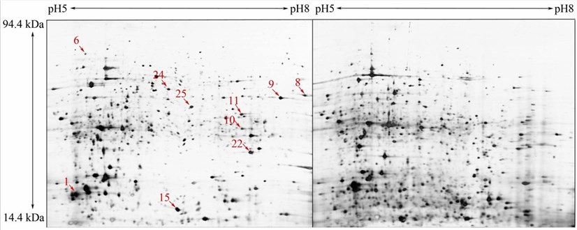 Fig.1 Two-dimensional sodium dodecyl sulfate-polyacrylamide gel electrophoresis (SDS-PAGE) gels of plant nematodes. (Fu HY, et al., 2014)
| Steps | Operation Methods |
| Protein Extraction | Nematodes are ground in a mortar with liquid nitrogen until a fine white powder is produced and then suspended with fresh weight ice-cold acetone containing TCA and dithiothreitol. The resulting protein-containing suspension is allowed to precipitate overnight at −20°C and then centrifuged. The pellet is rinsed three times with ice-cold acetone containing DTT at −20°C. Finally, the protein pellet is air-dried and dissolved in a lysis buffer solution. |
| IEF and SDS PAGE | Each sample containing protein in IEF buffer is loaded onto a 24 cm immobilized pH 5-8 gradient strip. Applying the appropriate procedure to focus the strip at 20°C. After focusing, proteins are reduced in the equilibration buffer containing DTT, followed by alkylation in a separate incubation for an additional 15 min in an equilibration buffer containing iodoacetamide. The strips are then transferred to 12.5% SDS PAGE gels for 2-DE using a gel system with SDS electrophoresis buffer. |
| Image Analysis | Protein spots are detected by silver staining. Stained gels are scanned and calibrated using the software. Detection and matching of the protein spots are facilitated with the use of the image software and re-evaluated by visual inspection. Spots that are expressed specifically in relatively high abundance are analyzed by mass methods. A positive identification must meet specific criteria. |
Lifeasible provides the detection of plant nematodes to our clients worldwide. With years of experience in biological services, our advanced platforms can help our clients solve various difficulties in plant protection. If you are interested in our services or have any questions, please feel free to contact us or make an online inquiry.
Reference
Get Latest Lifeasible News and Updates Directly to Your Inbox