Based on Lifeasible's advanced instrumentation and technical personnel, we can provide our clients with the most advanced liposome analysis services. We specialize in providing a full range of integrated services, including custom synthesis and characterization of liposomes, and can analyze liposomes' composition, shape, size and other parameters. Through a series of characterization tests, we can facilitate the development of liposomes and process optimization.
Liposomes are artificially prepared hollow microspheres, usually consisting of one or more phospholipid bilayers. Liposomes can be classified into various types according to particle size and a number of plasma membrane layers. Unilamellar liposomes consist of a single phospholipid bilayer encapsulated with good membrane properties and are easy to prepare. Oligomeric liposomes are induced by monolayer liposomes transformed and encapsulated by two layers of the plasma membrane. Multilamellar liposomes are encapsulated by several phospholipid bilayers. In contrast, multilamellar liposomes contain several small liposomes within one large liposome, which has the advantages of good encapsulation, high stability, and simple preparation. The spatial morphology of liposomes can be characterized by scanning electron microscopy, cryo-transmission electron microscopy, atomic force microscopy, and other instruments.
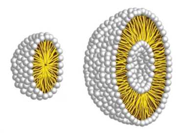
Lifeasible provides customers with liposome spatial morphology services to visually image liposome systems at the molecular level. The spatial morphology analysis provides liposome data, including size distribution, bilayer organization, and vesicle shape. We provide reliable instrumentation for spatial morphology analysis of liposomes and professional liposome solutions for our customers.
Our laboratory usually tests the structure of liposomes by atomic force microscopy. It is used not only to measure the forces at the molecular level and obtain images of the apparent shape but also to analyze elastic properties. It records the influence not only to show the shape properties of the sample but also by the softness of the sample.
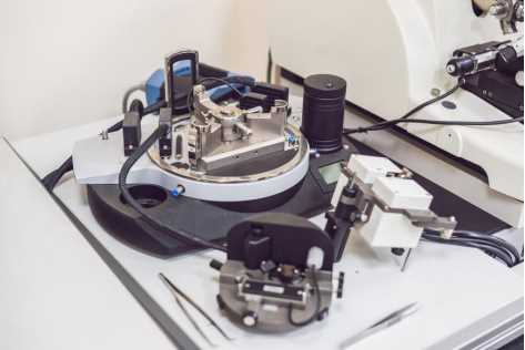
AFM can only scan samples adsorbed on substrates, not samples in suspension. It is more accurate when scanning liposomes in the liquid phase, but it is essential to ensure that the liposomes are adsorbed to the substrate. Consider chemical modification of the liposome and the substrate so that the liposome adsorbs to the substrate through chemical bonding, or consider adsorbing the liposome to the substrate through electrostatic forces between the liposome and the substrate. If multi-layer stacking is a concern, the liposomes can be diluted to a certain degree before smearing.
If you are interested in our services, or if the service you want is not listed, please feel free to contact us, and we will get back to you as soon as possible.
Get Latest Lifeasible News and Updates Directly to Your Inbox