Plant Telomere Specific Fluorescent Staining
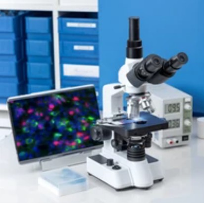
Fluorescent staining can provide high-resolution images with great specificity, allowing researchers to study the spatial distribution, interactions, and dynamics of different cellular components in living or fixed samples. Telomeres are repetitive DNA sequences at the ends of chromosomes that shorten with each cell division. Fluorescent staining of plant telomeres is a method used to visualize and study the structure of plant telomeres. In addition, fluorescent dyes can be used in various applications, such as flow cytometry, immunofluorescence, and fluorescent in situ hybridization (FISH), which have critical applications in research.
Lifeasible stands out in plant telomere-specific fluorescent staining due to its commitment to innovation, reliability, and customer satisfaction. With its expertise in telomere research and extensive range of products and services, we offer invaluable support to researchers in studying plant telomeres.
Highly Specific Telomere Staining Probes
- Lifeasible has pioneered the development of novel telomere-specific staining probes that offer enhanced specificity and sensitivity. These probes are designed to bind specifically to telomeric DNA sequences, allowing for selective visualization and quantification of telomeres.
- Our fluorescently labeled probes, such as FAM, Cy3, and Cy5, are used to label telomeric DNA, enabling their detection under a fluorescence microscope. These probes can be applied to various plant species, making them a valuable tool for researchers in the field.
Customized Staining Protocols
- Plant telomere-specific fluorescent staining requires optimized staining protocols to achieve accurate and reproducible results.
- We offer customized staining protocols to meet specific experimental requirements of scientific research, including variations in fixation methods, probe concentrations, and staining durations. Different plant species and tissues may require variations in staining conditions to ensure the successful detection of telomeres.
- By working closely with researchers and understanding their specific needs, we ensure that their staining protocols yield consistent and high-quality results. These protocols can be easily integrated into existing laboratory workflows, saving valuable time and effort for researchers.
Advanced Image Analysis Tools
- Accurate and comprehensive analysis of telomere staining images is crucial for extracting meaningful data from plant telomere-specific fluorescent studies.
- We offer advanced image analysis tools that facilitate precise quantification and characterization of telomeres, providing valuable insights into telomere length, distribution, and localization within plant cells. Our advanced algorithms ensure reliable and reproducible results, even in complex or noisy images. Our tools enable us to analyze large datasets efficiently and extract meaningful statistical information.
Lifeasible's highly specific telomere staining probes, customized staining protocols, and advanced image analysis tools provide researchers with the necessary resources to conduct meaningful and impactful telomere research. If you are interested in our services or have some questions, please feel free to contact us or make an online inquiry.
For research or industrial raw materials, not for personal medical use!

