Synthesis and Secretion Analysis of Calreticulin in Plant Nematodes
Calreticulin is a highly conserved and functionally rich protein family which widely exists in higher organisms and belongs to the endoplasmic reticulum retention protein family. It usually contains 3 major internal domains, consisting of a globular N domain and two Ca2+-binding domains. As reported by NCBI, the similarity of calreticulin between Meloidogyne enterolobii and Meloidogyne incognita is high, and it is also high with calreticulin from cereal cyst nematode, pine wood nematode, banana perforating nematode, rice dry tip nematode, and potato rot stem nematode.
Lifeasible offers comprehensive services covering a wide range of cutting-edge technologies to advance your projects. Our scientists have developed a series of innovative solutions to help analyze the synthesis and secretion of calreticulin in plant nematodes.
Calreticulin Synthesis Sites in Plant Nematodes
- The calreticulin Mi-CRT of the Meloidogyne incognita, which is an effector protein that inhibits host defense. It is mainly expressed in the sub ventral esophageal glands of the second instar nematode before infection and in the dorsal esophageal glands of the parasitic stage, indicating that it is mainly synthesized in the esophageal glands.
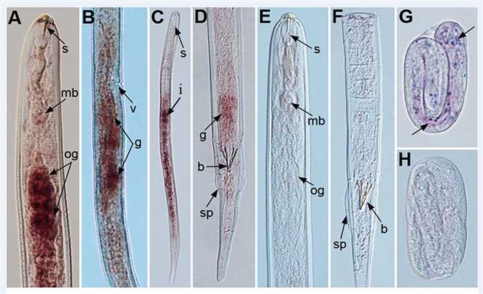 Fig.1 Tissue localization of Rs-crt mRNA via in situ hybridization. (Li Y et al., 2015)
- Lifeasible provides analysis of calreticulin synthesis sites in plant nematodes, including esophageal glands, gonads, intestines, eggs, etc. Several techniques, including in situ hybridization, are used to study the synthesis sites of calreticulin.
Developmental Expression of Calreticulin at Different Instar Stages
- The life cycle of sessile endophytic nematodes is usually divided into six stages, namely egg (the first instar larva), the second stage before infection (J2), the second stage after infection (ParJ2), the third stage (J3), the fourth stage (J4) and the adult stage (adult). After invading root tissue, J2 instar larvae establish permanent feeding sites within the root, thereby inducing the formation of syncytia and giant cells in root cells.
- Based on the biotechnology, including qRT-PCR and in situ hybridization, we provide developmental expression analysis of calreticulin at different instar stages from the egg to the adult stage.
Localization of Calreticulin in Plant Cells
- When plant nematodes infect their hosts, they use the oral needle to secrete effector proteins into host plants to change the structure and function of plant cells.
- We provide localization analysis of calreticulin in plant cells. Using immunohistochemical and subcellular localization techniques, calreticulin signals are detected in the cell walls, feeding sites, cytoplasm, endoplasmic reticulum, and Golgi apparatus of plant cells.
Lifeasible has extensive experience and expertise in plant science. We are committed to providing you with timely and high-quality deliverables. At the same time, we guarantee the cost-effectiveness, completeness, and simplicity of the report. If you are interested in our services or have any questions, please feel free to contact us or make an online inquiry.
Reference
- Li Y, et al. (2015). "A Nematode Calreticulin, Rs-CRT, Is a Key Effector in Reproduction and Pathogenicity of Radopholus similis." PLoS One.10 (6), e0129351.
For research or industrial raw materials, not for personal medical use!