Functional Analysis of the ER Body
ER body is a spindle-shaped and large structure, about 10 μm in length and 1 μm in width. Its main component is PYK10, a glucosidase with endoplasmic reticulum resident signal KDEL at the C terminus. In the event of injury, stress, or pathogen infection, it can react with the matrix to generate toxins and play a defensive role. In addition, the ER body also contains two stress-induced proteinases, RD21, and VPE.
Lifeasible provides functional analysis of ER body, which is responsible for storing and transporting hydrolytic enzymes, such as glucosidase and proteinases, to help our customers worldwide in the field of plant science. Our experts will answer your questions comprehensively and carefully and help you solve the problems in your studies. We guarantee satisfied and reliable results for our customers all over the world.
ER Body Stores Glucosidase
- ER body can store and transport PYK10, a member of the β-glucosidase (BGLU) family and a major component of the constitutive ER body.
- It has been reported that PYK10 mRNA is delivered to specific regions of the endoplasmic reticulum (ER), which promotes the concentration of PYK10 and the formation of ER body, or it may be that a large amount of PYK10 forms aggregates in the ER lumen and promotes the formation of ER body.
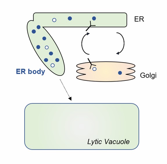 Fig.1. ER-derived compartments for hydrolytic enzyme storage.
- Lifeasible helps our customers study this process through the following methods, including the development of transgenic plants, wounding treatment, counting ER bodies, immunoblot analysis, immunoelectron microscopy, and phylogenetic.
- We use confocal microscopy to observe the number of ER bodies. The expression levels of PYK10-related genes are analyzed by qRT-PCR. In addition, the specific proteins that can induce ER bodies are detected by immunoblotting analysis.
- Furthermore, we provide services to study the molecular mechanisms that regulate ER body formation, including mapping the gene locus, determination of the transcriptional start site and 3' untranslated region of the gene, plasmid construction, preparation for specific antibodies, immunoblot analysis and so on.
ER Body Stores Proteinases
- In addition, the ER body also contains two Cys proteinases, RD21, and VPE. Both RD21 and VPE are vacuolar proteinases that are induced by environmental stresses.
- We provide experimental protocols to study this function of the MAG2 body, including the transformation of plants, ultrastructural analyses, microscopy, and subcellular fractionation.
Lifeasible offers functional analysis of the ER body with customized strategies and design. Our advanced technical platforms help our clients solve the problems they may encounter in research based on decades of experience. If you are interested in our services or have any questions, please feel free to contact us or make an online inquiry.
For research or industrial raw materials, not for personal medical use!

