Lifeasible offers a wide range of characterization tests for liposomes based on available equipment and technologies to facilitate product and process development of liposomes. lifeasible evaluates the permeability of liposomes through fluorescence technology, which allows us to determine the flux and rate of penetration of encapsulated small molecules.

Liposomes are synthetic spherical vesicles consisting of one or several lipid bilayers surrounding an internal water nucleus with particle sizes ranging from 20 nm to a few microns. Their composition usually includes natural or synthetic phospholipids. It may also contain sterols (most common cholesterol) and other lipid species (e.g., sphingomyelin, cardiolipin, etc.). The lipid bilayer sets up a hydrophobic barrier responsible for the selective transport of molecules across the membrane, a property known as membrane permeability. Membrane permeability is a measure of the flux or rate at which solutes cross the lipid bilayer to enter the aqueous compartment on the other side of the membrane. This is a critical biological function because transport across biological membranes involves many cellular processes considering cell viability. The molecular transport mechanisms of substances across biological membranes fall into two main categories: passive and active transport. Fluorescence techniques have proven to be very effective in monitoring the permeability of liposomal membranes and demonstrating the permeation of a large number of membrane-active agents.
Lifeasible offers customers the ability to test liposome permeability through fluorescence techniques, including fluorescence spectroscopy using self-burst probes, dye pairs or lanthanide cation/ligand pairs, and confocal fluorescence microscopy using single or multiple fluorophores.
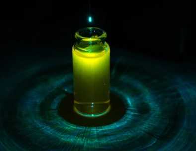
Fluorescent agents are encapsulated in liposomal aqueous compartments at self-bursting concentrations; membrane permeability is assessed by releasing them from the liposome over time. As fluorescent probes are released from liposomes, an increase in fluorescent signal correlates with increased permeability of the liposomal membrane. The most commonly used self-bursting probes are calcein, carboxyfluorescein, and sulforhodamine B. The kinetics of dye release can determine the permeability of liposomal membranes.
Vesicle leakage can be monitored using a pair of two reagents: the fluorophore and its bursting agent. Co-encapsulation of both reagents in the same liposomal core produces a fluorescence burst. The release of the fluorophore from the vesicle and its subsequent dilution is measured by the increase in fluorescence.
Dependent on the co-encapsulation of weakly fluorescent lanthanide cations and fluorescence enhancer ligands in the same liposome batch. This yields high fluorescence intensity due to ligand chelation of the cation. Therefore, releasing the cation/ligand pair from the liposome into a medium containing another chelator, such as ethylenediaminetetraacetic acid (EDTA), will result in reduced fluorescence due to dissociation of the complex.
A blank GUV (without any labeling) is incubated with a solution containing a fluorescent label and test agent. The influx of fluorescent dye within the vesicles is then monitored by time-lapse confocal microscopy. The stability of the permeabilization process can also be tested by adding another fluorescent agent to the vesicles and taking additional photographs to see if the permeabilization is temporary.
If you are interested in our services and products, please do not hesitate to contact us anytime.
Get Latest Lifeasible News and Updates Directly to Your Inbox