Analysis of Feeding Sites in Host Roots

Analysis of Feeding Sites in Host Roots

Plant nematodes are devastating pests and cause considerable yield losses in many crop plants. A large part of this damage is inflicted by the sedentary endoparasite nematodes, which include two major groups commonly known as a cyst (Heterodera spp, Globodera spp) and root-knot nematodes (Meloidogyne spp). Plant nematodes induce highly specialized feeding sites inside the roots of their host that, once established, support the growth and development of the parasite throughout their longer-term interaction. They share a sedentary lifestyle and induce feeding cells intimately associated with plant vascular tissue.

Lifeasible makes full use of our specialized libraries, selection strategies, and maturation techniques to analyze feeding sites and host penetration of plant nematodes. ​With advanced technology and experienced staff, we provide comprehensive services for global clients to support their research.

Root Knot Galls and Vascularization of Feeding Sites

  • After hatching from eggs in the soil, second-stage juveniles penetrate host root cells near the tip and follow an intercellular migratory path in the cell wall compartment to the zone containing differentiating vascular cells. Here, they select host target cells and initiate reprogramming events that result in the conversion of these target cells into giant cells.
  • Schematic representation of the root-knot nematode infestation life-cycle.
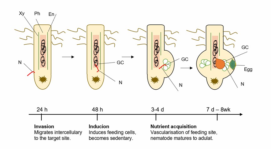
Fig.1 Schematic representation of the root-knot nematode infestation life-cycle.

  • Lifeasible provides analysis services for root knot galls and vascularization of feeding sites, including giant cells at a single feeding site and their connection with plant vasculature. In addition to the induction of giant cells, we help analyze other striking features of root nodules, which is the de novo formation of an extensive network of xylem and phloem vascular tissue that envelops giant cell clusters.

The De Novo Formation of Xylem

  • Xylem elements are readily identifiable in histological sections of feeding sites by their often-thicker cell walls and lignified secondary wall depositions.
  • We provide an examination of the giant cell complex depending on the three-dimensions; an extensive network of xylem cells can be found either surrounding the giant cells or localized to distinct regions along the giant cell border. In addition, we help customers to analyze the characteristics of xylem cells that differentiate in galls surrounding giant cells, as well as the underlying and controlling mechanisms of xylem cell formation and differentiation.

The De Novo Formation of Phloem

  • Extensive formation of phloem is induced around giant cells. In some regions, only phloem cells and very few other cell types are found directly adjacent to giant cells.
  • We provide an examination of phloem cells in root knots through a combination of fluorescence microscopy and immunohistochemistry. At the same time, we help to explore the mechanism by which the phloem embedded with giant cells does not contain typical companion cells.

Lifeasible offers professional services to meet your research needs. ​With years of experience in plant sciences, our advanced platforms can help our clients solve various difficulties and conduct research. If you are interested in our services or have any questions, please feel free to contact us or make an online inquiry.

Our products/services are For Research Use Only. Not For Clinical Use!
Online Inquiry