Observation of Endoplasmic Reticulum Structure in Plants
The endoplasmic reticulum (ER) is a membrane that performs many essential functions in a eukaryotic cell. It forms a 3-D lace-like network consisting of continuous tubules and flat lamellae, whose motility can be observed in living cells as ER structures in tubular flow cytoplasmic chains or tubular structures and lamellar plates. These changes include the movement of the tubules as well as their growth and contraction.
Lifeasible provides observation services of endoplasmic reticulum in plants to help our customers worldwide in plant scientific research. Our platform is equipped with cutting-edge facilities and professional experts to support research. Here, we provide various services according to customers' demands.
Observation of Endoplasmic Reticulum with Confocal Microscopy
- Among contemporary image acquisition techniques, confocal microscopy is particularly useful because it can perfectly obtain thin serial optical digital images of sections within a registered stack of thick specimens and 3-D image data in a large number of biological samples, including living specimens.
- The endoplasmic reticulum, which consists of tubules and lamellae, is detected by the local minimum threshold of the image intensity histogram. The area density of the ER layers of the cell epidermis is obtained by dividing the area of the pixels in the ER by the area of the region of interest. The length density of the tubules in the cortical layer of the cells is further estimated.
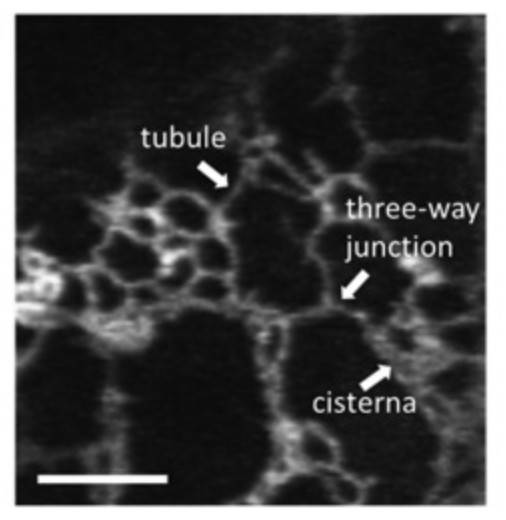 Fig.1 Confocal microscopic image of the cortical ER region of tobacco leaf epidermal cells. (Brandizzi F., 2021)
Observation of Endoplasmic Reticulum with Fluorescent Dyes
- In contrast to mitochondria and lysosomes, the ER is not readily visible under light microscopy in live or fixed cells, making the cellular distribution of the ER challenging to study.
- Lifeasible provides a fast and simple process for localizing a structure that appears to be the ER both in live cells and glutaraldehyde-fixed cells by fluorescent microscopy.
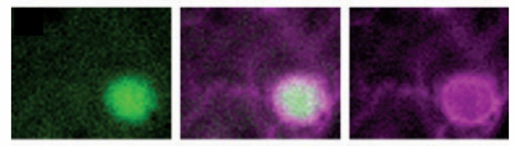 Fig.2 The bright YFP label is completely surrounded by the ER marker. (Nelson BK, et al., 2007)
- A series of fluorescent organelle markers are generated based on the perfect targeting sequence, which can be used for colocalization studies. This organelle marker set contains indicators for ER, Golgi, tonoplast, peroxisome, mitochondria, plastid, and plasma membrane.
- Based on the above principle, we select four different fluorescent proteins, including green, cyan, yellow or red, generated in two different binary plasmids, respectively for kanamycin or glucose phosphonate to allow for flexible combination. The labeled organelles show a characteristic morphology and can be used for their positive identification.
Lifeasible has a long-term commitment to the development and application of plant endoplasmic reticulum. We are pleased to use our extensive experience and advanced platform to provide satisfactory service and qualified products to meet the needs of our customers. If you are interested in our services or have any questions, please feel free to contact us or make an online inquiry.
References
- Brandizzi F. (2021). "Maintaining the structural and functional homeostasis of the plant endoplasmic reticulum". Dev Cell. 56 (7), 919-932.
- Nelson BK, et al. (2007). "A multicolored set of in vivo organelle markers for co-localization studies in Arabidopsis and other plants". Plant J. 51 (6), 1126-36.
For research or industrial raw materials, not for personal medical use!