Isozymes, as a class of proteins, exist widely in organisms. The main separation methods include electrophoresis, chromatography, enzymatic method, and immunological method. Enzymatic phenotyping is one of the first non-morphological based methods used for nematode identification. After the soluble protein is extracted from the plant nematode, the specific isoenzymes are stained, and the different nematode species can be identified by polyacrylamide gel electrophoresis analysis.
Lifeasible makes full use of our specialized libraries, selection strategies, and maturation techniques to optimize the stability and performance of plant protection. With advanced technology and experienced staff, we provide protocols for the detection of plant nematodes to global clients to support their research.
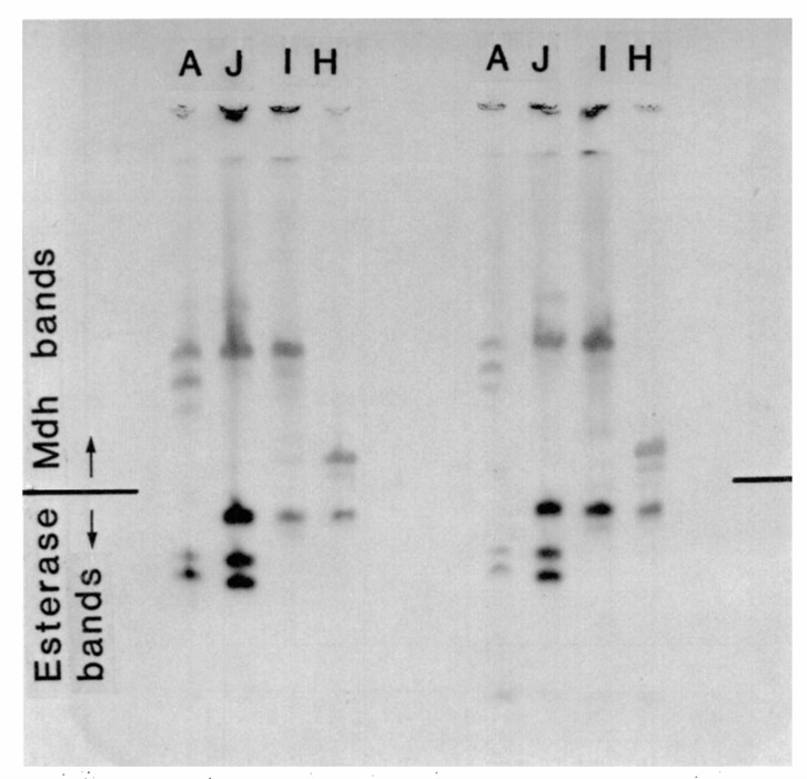 Fig.1 A polyacrylamide gel showing the esterase and malate dehydrogenase phenotypes of plant nematode species. (Esbenshade PR and Triantaphyllou AC., 1990)
| Steps | Operation Methods |
| Enzyme Solution Extraction of Plant Nematodes | Nematodes are separated by the funnel method, washed with distilled water, counted, and concentrated. The extract is added at 4°C, ground thoroughly in an ice bath, centrifuged for 15 min with 10000 g, and the supernatant is taken as an electrophoresis sample. |
| Making Glue | Pour the prepared separation glue into the glass plate immediately, and slowly pour the concentrated glue after the gel is polymerized. Then let the mold stand still for an hour, pour the electrode buffer into the electrophoresis tank, and prepare the sample. |
| Adding samples | 8 μL Sample is added to each well, and then one drop of bromophenol blue solution is added to the upper tank. |
| Electrophoresis | We perform the vertical polar polyacrylamide gel electrophoresis. The electrophoresis tank is placed horizontally in the refrigerator, maintaining the temperature at 4°C to keep the enzyme's activity. |
| Staining |
|
Lifeasible offers reliable protocols for the detection of plant nematodes to meet your research demands. With years of experience in plant science, our advanced platforms can help our clients solve various difficulties and conduct research. If you are interested in our services or have any questions, please feel free to contact us or make an online inquiry.
Reference
Get Latest Lifeasible News and Updates Directly to Your Inbox