Functional Analysis of the KDEL Vesicle
The diameter of KDEL vesicle (KV) is between 200 nm and 500 nm. After budding from the endoplasmic reticulum (ER), KV reaches the vacuole directly without passing through the Golgi. In plants, KV formation and vacuolar transport through KV are associated with the KDEL sequence of cysteine protease with KDEL signal. Most soluble ER resident proteins contain the KDEL or HDEL 4 peptide sequence at the C-terminus, called ER resident signal. The Golgi apparatus recognizes this signal and sends the resident proteins that have escaped back to the ER. Besides, the H / KDEL system is highly conserved in plants.
Lifeasible develops an advanced platform equipped with advanced instruments and professional staff for functional analysis of KDEL vesicles with a high standard. We customize featured services according to the customers' demand.
KDEL Vesicle Stores Cys Proteinase
- Sulfhydryl endopeptidase (SH-EP) is a cysteine proteinase belonging to Papain hydrolase. Its C terminus contains KDEL signal, related to the degradation of stored proteins in protein storage vacuoles. SH-EP has strong hydrolase activity and broad substrate specificity.
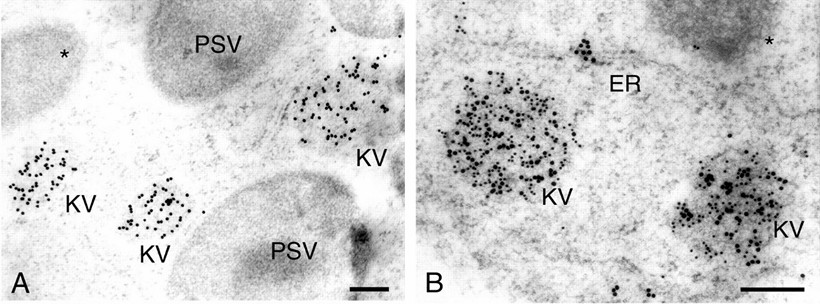 Fig.1. Electron photographs showing that KV is immunogold labeled with antibody to KDEL sequence. (Toyooka K, et al., 2000)
- Lifeasible helps our customers study this process through the following methods, including gel electrophoresis and immunoblotting, preparation of antibodies, immunocytochemistry and ultrastructural analysis, etc.
- We prepare transverse sections of plant organs and stain with toluidine blue to observe the pattern of protein storage in the cells. The localization of SH-EP in cell structure is observed by immunogold labeling plant cells with polyclonal antibody to SH-EP. Double immunogold staining is performed to distinguish localization in vesicles from PSV by polyclonal antibody against globulin and SH-EP.
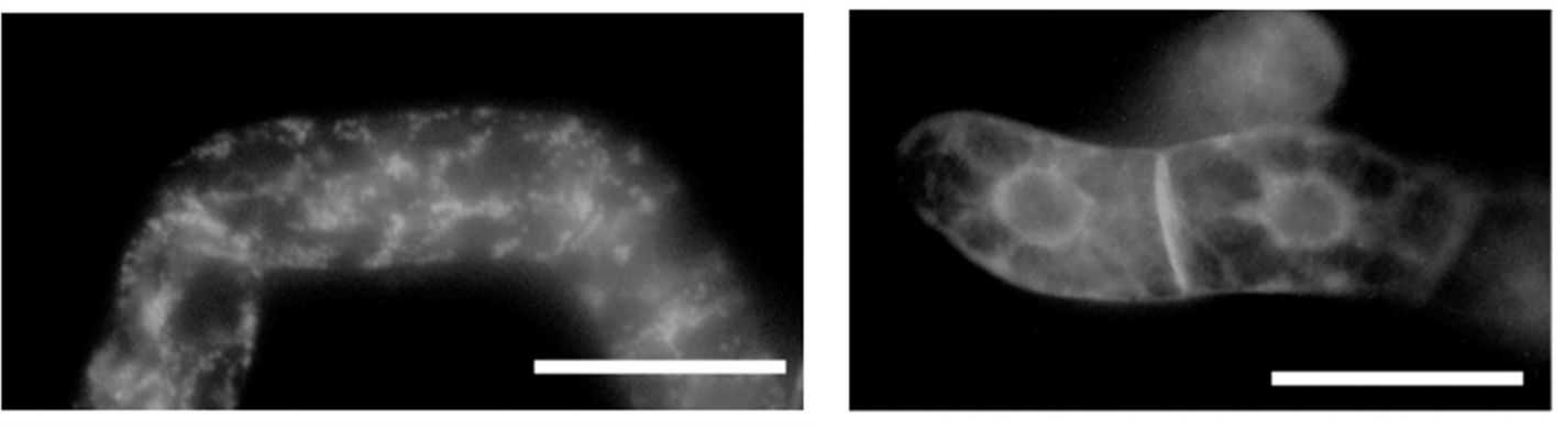 Fig.2. Intracellular localization of SP-GFP-SHEP or SP-GFP-SHEPΔKDEL in tobacco BY-2 cells. (Okamoto T, et al., 2003)
- In addition, the C-terminal KDEL sequence of KDEL tail cysteine protease is involved in the formation of KV.
- Lifeasible helps our customers study this process through the following methods, including preparation of transgenic plant cell expressing wild-type SH-EP or its KDEL deletion mutant (SH-EPΔKDEL), immunogold electron microscopy, etc.
- To understand the function of the KDEL sequence of SH-EP, we express wild-type SH-EP and SH-EPΔKDEL in plant cells and observe the formation of KV.
Lifeasible offers services covering functional analysis of the KDEL vesicle to meet your research demands. With years of experience in plant science, our professional platforms can help our clients solve various difficulties. If you are interested in our services or have any questions, please feel free to contact us or make an online inquiry.
References
- Toyooka K, et al. (2000). "Mass transport of proform of a KDEL-tailed cysteine proteinase (SH-EP) to protein storage vacuoles by endoplasmic reticulum-derived vesicle is involved in protein mobilization in germinating seeds." J Cell Biol. 148 (3), 453-64.
- Okamoto T, et al. (2003). "C-terminal KDEL sequence of a KDEL-tailed cysteine proteinase (sulfhydryl-endopeptidase) is involved in formation of KDEL vesicle and in efficient vacuolar transport of sulfhydryl-endopeptidase." Plant Physiol. 132 (4), 1892-900.
For research or industrial raw materials, not for personal medical use!

